 |
||
 MRI films
|
||
|
|
|
|||||||
| iSpine Discuss MRI films in the Main forums forums; When my symptoms first started I was a wreck, I could not process information well and was in such severe ... |
 |
|
|
LinkBack | Thread Tools | Display Modes |
 05-10-2009, 01:52 PM
|
|||
|
|||
|
When my symptoms first started I was a wreck, I could not process information well and was in such severe pain. When the first MRI's came back along with the first "you have a herniation but it's reall small", I decided to start finding my own information. Well, trying to find many MRI films to compare was very difficult. Google brings up a few, but the more you look, the more you find the same pictures over and over.
So, here are some of mine. The left is the sagittal view where you can clearly see the herniation. The bright spot in it is evidence for an annular tear. The right side is the axial view (looking from top down) of the C5-6 disc. You can see the bright spot in this view as well. 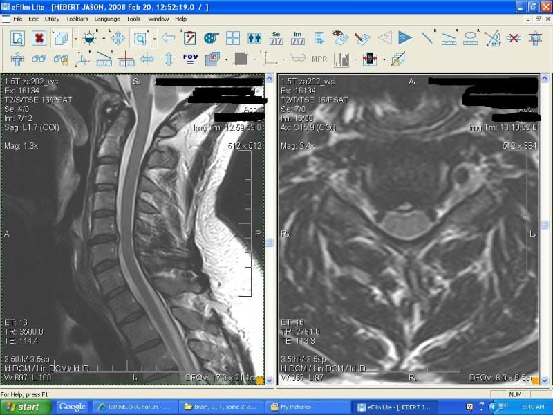 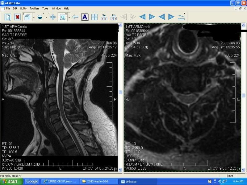
__________________
Chiari 1 malformation - successful surgery 1-22-09 C5-6 herniation (extrusion) with moderate central canal stenosis and bilateral foraminal stenosis. Prodisc-C @ C5-6 surgery on 5/28/09 VATS thoracic fusion @ T3-4 and T6-7 on 9/11/09 Fusion w/cage @ C7-T1 on 11/12/09 
|
|
05-26-2009, 04:14 PM
|
|||
|
|||
|
Even though my surgery is only two days away, I went ahead and requested a set of Flexion Extension MRI's. Although I do not have the report, here are some pics.
__________________
Chiari 1 malformation - successful surgery 1-22-09 C5-6 herniation (extrusion) with moderate central canal stenosis and bilateral foraminal stenosis. Prodisc-C @ C5-6 surgery on 5/28/09 VATS thoracic fusion @ T3-4 and T6-7 on 9/11/09 Fusion w/cage @ C7-T1 on 11/12/09
|
|
05-27-2009, 04:18 AM
|
|||
|
|||
|
If I did not know better, I would think that those were my films!
Even though I had a clear bulge at C5/C6 and impingement and displacement of the cord, I should have had a discogram. As it turns out C6/C7 and C4/C5 have some major tears and fissures, without having any bulges. Know that I have had a discogram, it is evident that I should have had a 2 level ADR. Good luck with your surgery...keep us posted... pun intended 
__________________
MVA 2005 - impinged and displaced cord at C5/C6 Prodisc C5/C6 2006 Germany - Dr. Bertagnoli C6/C7 no bulge, just tears and fissures. Multiple ministrokes 2009 prevents ADR surgery As of 10/2010 no relief on radicular C7 Trying to arrange C6/C7 prodisc FDA has not approved for 2nd Levels on cervical. Headed to Germany as soon as I am cleared for surgery.
|
|
06-07-2009, 03:04 PM
|
|||
|
|||
|
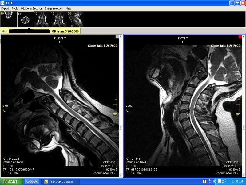
Here are another couple slices of my flexion extension MRI. I want to point out that these are weight bearing since I was sitting. The picture on the right is in a neutral position.
I also want to point out that I could not extend any more due to pain. If I would have been able to, I am sure that the herniations would have appeared larger. I am going to bold this because I think it is very important. This just proves that when you are in pain your body is telling you that there is something wrong. I think that in some situations, decreasing the pain without fixing the problem puts you at further risk of more injury. Also the two sets of pictures at the beginning of the thread are lay down traditional MRI's. It does not take an expert to see what traditional MRI's can miss and even weight bearing MRI's without flexion. Notice the difference in size of the herniation at T1-T2 between the extension position on the left and the neutral position on the right. Also note that the herniation at T3-4 appears smaller on the extension (left) MRI. When I try to alleviate the pain in my upper back I extend my neck like this, which in turn hurts my lower neck. I can't believe I have visual proof of my pain and I still get blown off. Both of these are wieght bearing. 
__________________
Chiari 1 malformation - successful surgery 1-22-09 C5-6 herniation (extrusion) with moderate central canal stenosis and bilateral foraminal stenosis. Prodisc-C @ C5-6 surgery on 5/28/09 VATS thoracic fusion @ T3-4 and T6-7 on 9/11/09 Fusion w/cage @ C7-T1 on 11/12/09 Last edited by jchebert1979; 06-07-2009 at 03:14 PM.
|
|
| Bookmarks |
|
|


 Linear Mode


